A radiologist with years of expertise could still scan an image in minutes or a few hours. But the ones who are in the beginning might need the help of an expert.
Delays in radiology mean risking a life.
These delays are due to the time-consuming process of scanning an image without the scope of any error. But still, some studies show that diagnostic error rates in radiology range from 3% to 5% globally. The percentage is less, but it’s a serious risk for hospitals and patients alike.
AI in radiology is the latest tech to spot these errors. These tools can spot diseases faster, often before symptoms even show up.
This blog will discuss the intersection of artificial intelligence and radiology, covering its history, working, benefits, and real-world use cases.
Let’s decode.
Brief History of Radiology & Medical Imaging
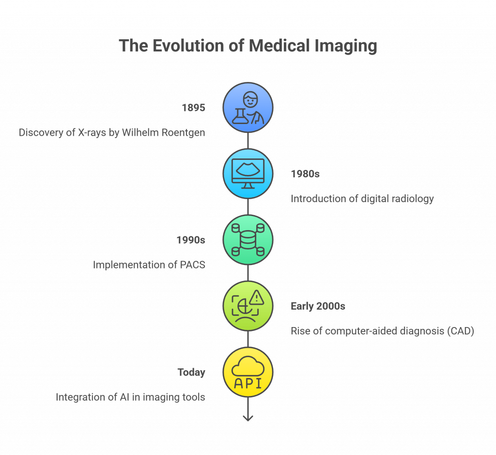
In 1895, Wilhelm Roentgen was experimenting with cathode ray tubes when he accidentally discovered X-rays. Within weeks, doctors started using them to look inside the human body. It was where modern radiology began.
For decades, imaging was film-based. Radiologists developed and read scans manually, which was slow and required high expertise. Then came digital radiology in the 1980s. At that time, scans could be stored, shared, and reviewed on computers.
The 1990s welcomed PACS – Picture Archiving and Communication Systems. This helped hospitals store massive volumes of imaging data securely and access it remotely. It laid the foundation for large-scale diagnostic workflows.
The early 2000s saw the rise of computer-aided diagnosis (CAD) tools. These were basic algorithms that flagged potential issues in mammograms or lung scans. But they lacked intelligence and needed a lot of human supervision.
Today, we are moving toward machine learning and AI. Imaging tools are becoming smarter, faster, and more accurate with the incorporation of AI.

Emergence of AI in Radiology
The path of AI and radiology is built on a series of scientific leaps:
- It was in 2010 when AI started its pace. Deep learning, a form of machine learning, became popular for its success in image recognition. This mattered for radiology because medical scans are, essentially, complex images filled with data.
- Thereafter, in 2016, Google’s DeepMind brought out a model in the market that could detect diabetic retinopathy, which is a serious eye condition. What’s interesting is that it matched the accuracy of high-level expert doctors. For the first time, the doctors started believing that AI could make safe medical decisions.
- By 2018, the FDA approved the first AI software that could analyze chest X-rays without a human radiologist. That tool, built by Zebra Medical, showed how AI could become a trusted second opinion, especially in highly crowded hospitals. This also opened doors for a new generation of healthcare software focused on diagnostic accuracy and speed.
- Between 2020 and 2025, AI boomed. A wide number of startup organizations launched their AI tools for different types of scans, be it X-rays, MRIs, or CTs.
- As of 2025, over 400 AI-based medical devices have FDA or CE approvals. That means they meet strict quality and safety standards.
- For businesses in healthcare, these milestones show that AI in medical imaging has come a long way, and that, too, with high reliability.

deepcOS
Starting Price
Price on Request
How AI Works in Radiology?
AI’s role in radiology is to support human experts, making their tasks easier.
The process starts with machine learning and deep learning. These AI subsets train computers to read and understand images by providing them the annotated medical images. These images have already been declared by an expert radiologist which identify conditions like fractures, tumors, or infections.

Here’s a step-by-step look at how AI analyzes a medical scan:
- Image Acquisition: A patient undergoes a scan such as an X-ray, CT, or MRI. The raw image is captured by the imaging machine.
- Preprocessing: The AI system makes the images clearer to read. It removes noise, adjusts brightness and contrast, and enhances important features. Preprocessing ensures the AI sees a clear and standardized image for analysis.
- AI Analysis: The cleaned image is fed into the AI model. The model scans every pixel, comparing patterns that it has learned while training. It identifies potential abnormalities like nodules, fractures, or unusual tissue density.
- Highlighting Findings: The AI generates output highlighting areas of concern. Often, it produces heatmaps or bounding boxes over suspicious regions, showing exactly where it detected anomalies.
- Radiologist Review: A trained radiologist reviews both the original and AI scans. The AI acts like a second pair of eyes, helping the radiologist confirm or rule out issues more quickly.
- Report Generation: Based on this combined review, the radiologist creates the final diagnostic report that finally goes to the clinician or patient.
- Feedback Loop: In some advanced systems, radiologists can also provide feedback on AI suggestions. This further helps in improving the AI model over time.
Key Benefits of AI in Radiology
Here are some benefits of Artificial Intelligence in radiology:
- The first advantage is the speed of the AI. AI provides quicker interpretation of scans. Its algorithms are able to analyze images within seconds, accelerating the diagnosis. Radiologists can thus make faster decisions for any treatment.
- Second, it provides a precise diagnosis. Research indicates that AI has the ability to identify abnormalities as accurately as humans or even more so. This minimizes false positive results and missed diagnoses.
- Another advantage AI offers is the early detection of diseases. AI can detect minute symptoms that could sometimes escape the human eye. This helps in treating the specific disease early, increasing the survival rates in diseases such as cancer and stroke.
- AI Automation of routine jobs can reduce radiologists’ burnout. They can concentrate on more complicated cases that require human intelligence.
- Lastly, AI provides smooth second opinions. Where radiologists encounter doubtful cases, AI offers a second opinion that they can trust. This enhances confidence in diagnoses.
Real-World Applications of AI in Radiology
AI is being applied across key radiology domains. Here are a few use cases to understand AI’s importance.
1. Pulmonology: Chest X-rays & Lung Disease
A chest X-ray is the most common medical scan. But it’s difficult and takes ample time to manually detect conditions like pneumonia, tuberculosis, or lung nodules.
Many tools today, like Qure.ai qXR, use AI to read chest X-rays and detect up to 29 lung conditions, including TB and COVID-19. It’s already being used widely in public health programs across Asia and Africa.
Similarly, CAD4TB is specialized for tuberculosis screening. CAD4TB holds CE certification that meets international safety and quality standards.

Qure AI
Starting Price
Price on Request
2. Oncology: Breast Cancer Detection
Breast cancer takes the lives of many worldwide. But survival rates can be improved if the disease is detected early. Dense breast tissue, subtle lesions, and variability between radiologists can cause missed diagnoses or false alarms.
Lunit INSIGHT MMG uses AI to detect suspicious lesions and classify them by risk level. It also calculates breast density scores, which are an important factor in assessing cancer risk.
3. Orthopedics: Fracture Detection
Bone fractures generally require emergency treatment. In the hustle, it becomes difficult for radiologists to examine large volumes of X-rays, and that too under time pressure. Many complications can occur if there is a slight delay in fracture diagnoses.
AZmed Rayvolve is a tool that takes the help of AI and detects fractures from X-ray images within seconds. The tool highlights fracture sites with clear visual markers, helping clinicians confirm findings faster.
4. Brain & Spine Imaging
Neurological disorders and spinal injuries often require detailed imaging and careful interpretation. Lesions, aneurysms, multiple sclerosis plaques, and vertebral fractures can be subtle and difficult to detect.
mdbrain provides volumetric analysis of brain MRIs. For spine imaging, IB Lab FLAMINGO uses AI to identify vertebral compression fractures from CT scans. This supports orthopedists and neurologists by providing fast and reliable detection.
5. Liver & Biliary Imaging
The liver and biliary system are complex, and imaging these organs can be challenging. But AI now enhances this imaging with 3D biliary mapping and detailed tissue characterization.
Perspectum MRCP+ applies AI to MRI scans to create accurate 3D maps of the bile ducts. This level of detail helps hepatologists and radiologists tailor treatment plans and monitor disease progression effectively.
Conclusion
AI in radiology comes with challenges. But the benefits are too big to ignore.
AI speeds up diagnosis with improved accuracy. Studies show that AI tools can cut diagnostic errors by up to 30%. That’s a major win for patient safety.
Many healthcare businesses embrace AI as it has resulted in better operations and patient services.
To deliver top-tier care, now is the high time to invest in AI imaging as the future of healthcare lies in it.
Mehlika Bathla is a passionate content writer who turns complex tech ideas into simple words. For over 4 years in the tech industry, she has crafted helpful content like technical documentation, user guides, UX content, website content, social media copies, and SEO-driven blogs. She is highly skilled in... Read more


